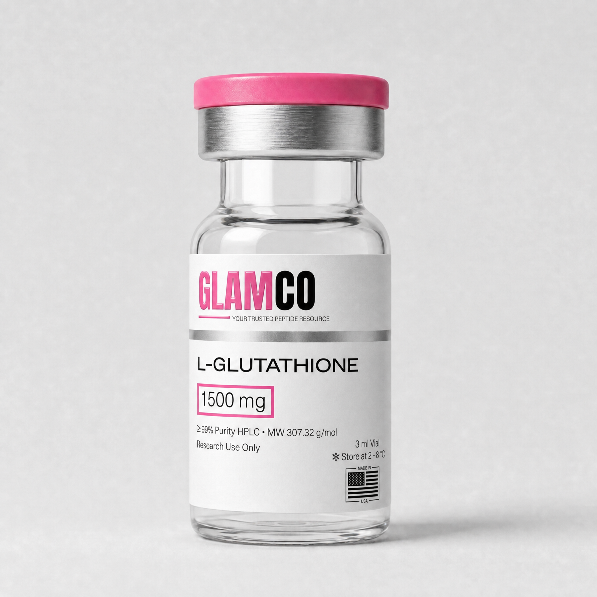
L-Glutathione
L-Glutathione (reduced GSH) lyophilized intracellular tripeptide antioxidant (γ-Glu-Cys-Gly) for in vitro redox-biology research.
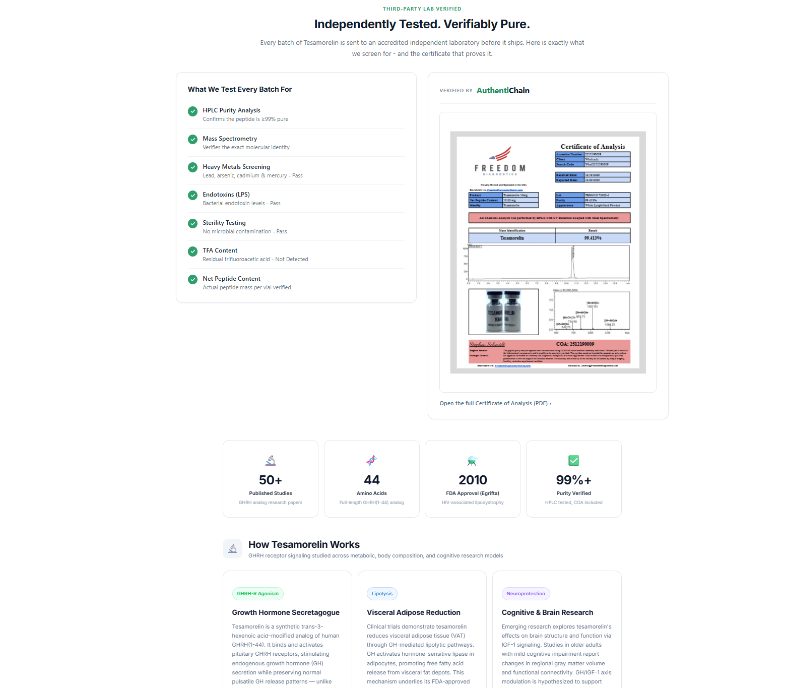
Independently Tested. Verifiably Pure.
Every batch of L-Glutathione is sent to an accredited independent laboratory before it ships. Here is exactly what we screen for - and the certificate that proves it.
What We Test Every Batch For
How L-Glutathione Works
Three converging redox pathways studied in cell-culture and biochemical research models
ROS Scavenging & Redox Cycling (GSH↔GSSG)
The free cysteine thiol (-SH) on GSH donates electrons to reactive oxygen species, including hydrogen peroxide and lipid peroxides, generating oxidized glutathione disulfide (GSSG). Glutathione reductase regenerates GSH from GSSG at the expense of NADPH, maintaining the cellular GSH:GSSG ratio that defines redox potential (Lu, 2013).
- Direct quenching of hydroxyl, peroxyl, and peroxynitrite radicals
- Substrate for glutathione peroxidase (GPx) reducing H₂O₂ and ROOH
- NADPH-dependent recycling via glutathione reductase (GSR)
Conjugation via Glutathione S-Transferase
Glutathione S-transferases (GSTs) catalyze nucleophilic addition of the GSH thiol to electrophilic xenobiotics, drug metabolites, and lipid-derived aldehydes (4-HNE). The resulting GSH-conjugates enter the mercapturate pathway for renal/biliary export, a central Phase II detoxification route (Forman et al., 2009).
- Catalytic substrate for the GST enzyme superfamily
- Detoxifies electrophilic drug metabolites & xenobiotics
- Sequesters reactive aldehydes (4-HNE, MDA) in oxidative-stress models
Antioxidant Defense Pathways in Cell Models
GSH integrates with the wider antioxidant network: regenerating ascorbate and α-tocopherol, supporting peroxiredoxin/thioredoxin redox cycles via the Nrf2-driven gene program, and S-glutathionylating protein cysteines as a reversible post-translational redox switch (Aoyama et al., 2008).
- Regenerates oxidized vitamins C and E in vitro
- Couples to Nrf2/ARE-driven antioxidant gene expression
- Reversible protein S-glutathionylation as redox signaling switch
What Research Has Shown
Key preclinical findings from peer-reviewed glutathione literature
Research Applications
Primary preclinical areas of L-Glutathione investigation
Oxidative Stress Research
GSH is the dominant intracellular antioxidant in mammalian cells. Cell-culture models manipulate GSH levels (BSO depletion, exogenous GSH addition) to study ROS-driven apoptosis, ferroptosis, and lipid peroxidation, with the GSH:GSSG ratio serving as the canonical readout of cellular redox state.
Lu 2013 ↗Hepatic Detoxification Pathways
The liver maintains the highest GSH concentrations in the body and is the principal site of GST-mediated Phase II conjugation. Preclinical models use GSH-depletion (acetaminophen, CCl₄) to study drug-induced liver injury, while GSH precursors (NAC, GSH ethyl ester) are studied as rescue agents.
Forman et al. 2009 ↗Skin Pigmentation / Tyrosinase Research
In vitro and ex vivo melanocyte models report that GSH inhibits tyrosinase activity and shifts melanogenesis from eumelanin toward pheomelanin via thiol-mediated copper chelation, supporting GSH as a research tool compound in melanin-biology and skin-pigmentation cell models.
Sonthalia et al. 2017 ↗Neurodegeneration Preclinical Models
Brain GSH depletion is consistently reported in preclinical Parkinson's and Alzheimer's models. Neuronal GSH synthesis (via the EAAC1 cysteine transporter) and dopaminergic neuron vulnerability to oxidative stress are central research questions in cell-based and rodent neurodegeneration models.
Aoyama et al. 2008 ↗Compound Information
Technical specifications and analytical profile
Frequently Asked Questions
Common questions about L-Glutathione research parameters
Sources & References
Peer-reviewed publications and preclinical studies database